Friday, March 1, 2013
Stress, the Brain, & Chronic Pain

Patients with chronic or recurring pain know how stressful their conditions can become. New research suggests that there may be physiologic causes of such stress, mediated by changes in brain structure that make these persons even more vulnerable to stress and pain. While there were many limitations of the study, there are implications for the importance of stress-reduction techniques in helping to better manage chronic pain.
The study — conducted primarily by Canadian researchers at the Université de Montréal — included 16 patients with chronic back pain and a control group of 18 healthy subjects [Vachon-Presseau et al. 2013]. The research objective was to analyze relationships between 4 factors: 1) cortisol levels, which were determined via saliva samples collected during 7 days; 2) assessments of clinical pain reported by patients prior to brain scans (self-perception of pain); 3) the size of brain hippocampal regions measured using magnetic resonance imaging (MRI); and 4) brain activations assessed with functional MRI (fMRI) following application of a thermal pain stimulus.
The results, reported in the journal Brain, showed that patients with chronic pain generally have higher cortisol levels than healthy individuals. Cortisol, produced by the adrenal glands, is sometimes called the “stress hormone” since it is activated in reaction to stress. It is a steroid hormone that increases blood sugar, suppresses the immune system, and may decrease bone formation; so, an excess is deleterious to health.
Data analysis further revealed that higher cortisol also was associated with smaller hippocampal volume and stronger pain-evoked activity in the anterior parahippocampal gyrus, a region involved in anticipatory anxiety and associative learning. Brain activity in response to the painful stimulus, assessed during fMRI scanning, reflected the intensity of the patient’s current clinical pain condition.
The authors conclude that their findings support a “stress model of chronic pain,” suggesting that the sustained endocrine stress response observed in individuals with a smaller hippocampus induces changes in the function of the hippocampal complex, which in turn may contribute to increased and persistent pain states. Such findings imply that stress management interventions might be important adjunctive treatments in the management of chronic pain.
COMMENTARY: This study helps to elucidate understandings of the neurobiological mechanisms affecting chronic pain and the vital role of stress. It supports recent theories suggesting that chronic pain could be partly maintained by maladaptive neurophysiological responses to recurrent stressors.

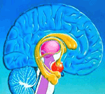
We have previously discussed how structural and functional alterations in the brains of persons with various chronic pain syndromes may influence the persistence and intensity (and possibly the initial development) of their conditions. See the series of pain-and-the-brain UPDATES [here]. This present study examines a possible role of the hippocampus, a component of the limbic system that plays important roles in the consolidation of information from short-term memory to long-term memory as well as spatial navigation. Its importance for influencing the experience of pain has been less well documented.
Vachon-Presseau and colleagues [2013] suggest that their findings “open up avenues for people who suffer from pain to find treatments that may decrease its impact and perhaps even prevent chronicity. To complement their medical treatment, pain sufferers can also work on their stress management and fear of pain by getting help from a psychologist and trying relaxation or meditation techniques.”
Indeed, nonpharmacologic modalities — eg, relaxation, meditation, biofeedback, cognitive behavioral therapy, yoga, massage, and other stress and anxiety reduction techniques — may be important adjuncts for effective pain management. For example, a prior UPDATE [here] described how research on meditation found that this approach favorably influenced how participants’ brains responded to pain. In particular, as a result of meditation, multiple limbic structures reacted to pain in ways that moderated subjects’ processing of painful stimuli and their perceptions of pain. Therefore, it makes sense that structural hippocampus changes found in this present experiment by Vachon-Presseau could have an impact.
At the same time, however, there were some important limitations of this present research. For one thing, sample sizes were quite small — 16 in the experimental group, 18 controls — which raises questions about statistical veracity (eg, random effects), whether participants were truly representative of the larger population in question, and external validity. Small sample sizes are typical of imaging studies in the pain field, but larger-scale trials needed to substantiate reliability of outcomes are rarely performed.
Furthermore, causation and temporality cannot be confirmed by this study. That is, which came first, stress and an undesirable cortisol response or chronic pain? Did alterations of brain structure precede chronic pain or develop in response to it? Lastly, it would be important to know if the cortisol and brain-structure aberrations can be reversed by therapeutic approaches — whether pharmacologic or nonpharmacologic — and the ultimate effect of this remediation on chronic pain.
REFERENCE: Vachon-Presseau E, Roy M, Martel M-O, et al. The stress model of chronic pain: evidence from basal cortisol and hippocampal structure and function in humans. Brain. 2013;136(3):815-827 [abstract].